MISTac Is an 8 mm Tactile Finger Built to Fit a Surgical Trocar
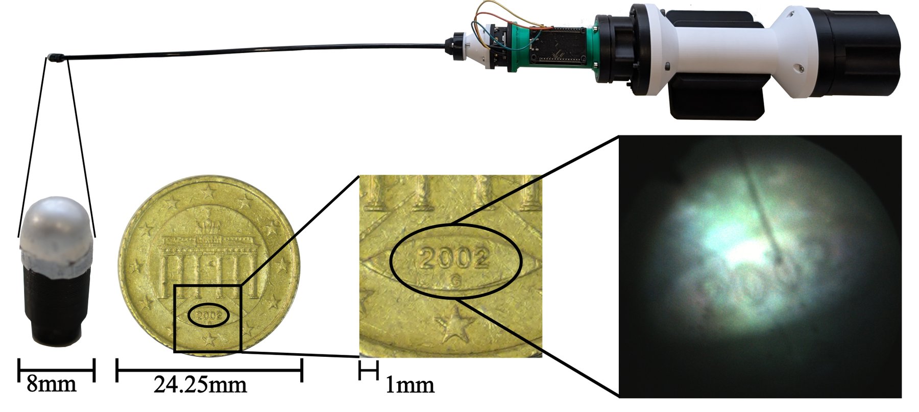
Robin Koch, Roberto Calandra, and collaborators at TU Dresden, Stanford, KIT, and Dresden’s surgical hospital posted MISTac on August 14: a vision-based tactile sensor whose replaceable tip is 8 mm across, sized for the trocars used in minimally invasive surgery.
They open-sourced the CAD, BOM, and lighting firmware at github.com/lasr-lab/mistac. The build guide is still marked “coming soon.”
DIGIT Pinki, shrunk for a port
MISTac borrows the DIGIT Pinki idea: keep the camera and LEDs at the proximal end, and pipe the image through a coherent fiber bundle. Pinki’s tip is 15 mm. That is too fat for typical 8 mm surgical ports.
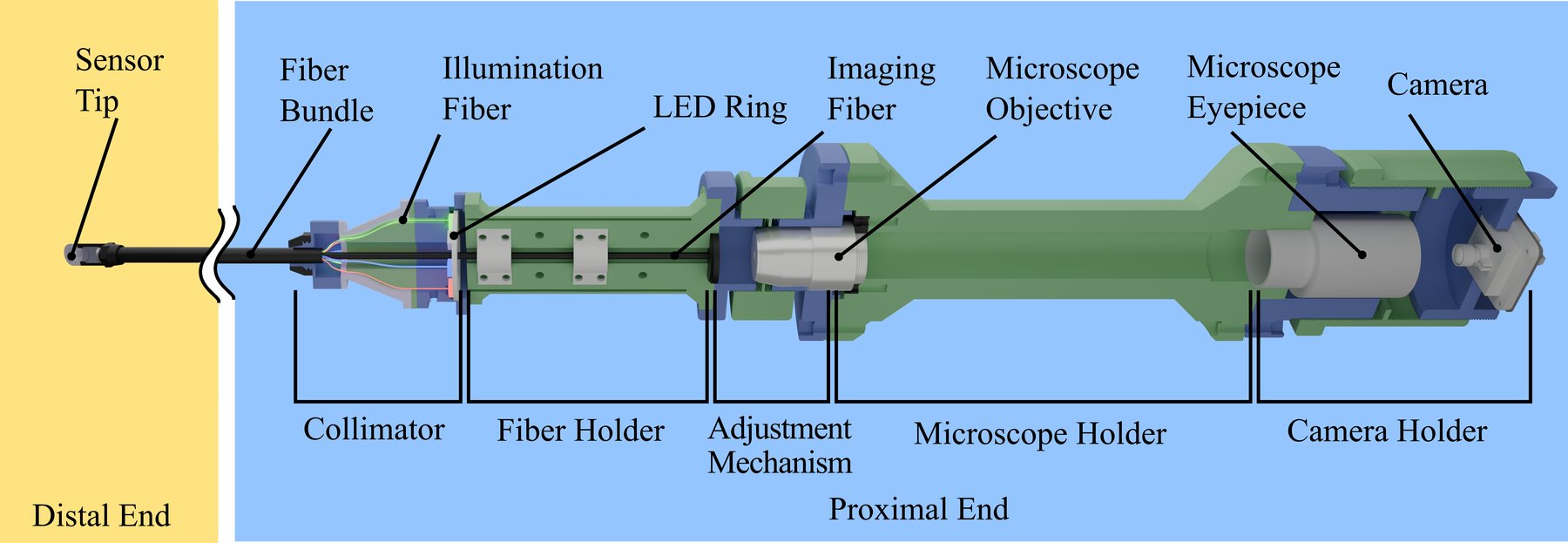
The new tip is an elliptical silicone gel, 12 mm long, in a 3D-printed cartridge that screws onto the fiber with a nut. Swap it between procedures. The proximal case is four printed subassemblies: collimator, fiber holder, microscope holder, camera holder. An Adafruit NeoPixel ring plus an ESP32 can change RGB lighting in software.
Measured numbers from their bench:
- Optical resolution: 176.68 µm (USAF 1951)
- Tactile resolution: 250 µm on a milled aluminum block
- Force RMSE: 24.3 ± 0.9 mN with a ResNet-18, against a BotaSys sensor whose own noise is about 20 mN
They actually put it through a trocar
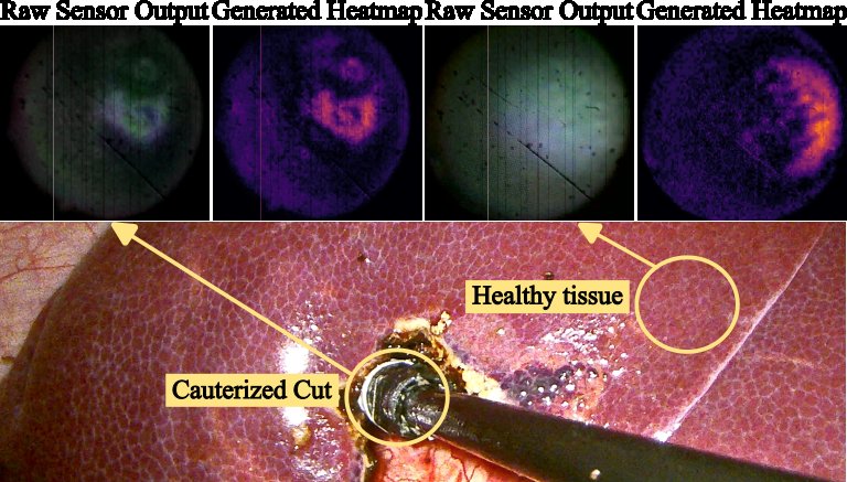
The medical test used an older camera (Arducam B0268) on n=2 sedated pigs, under approval TVV43/2023. A surgeon palpated through one port while an assistant ran the endoscope through another. They recorded 40 minutes at 30 fps.
A ResNet-18 plus a small causal Transformer then classified four tissues: liver, cauterized liver cut, spleen, and colon. Leave-one-out accuracy was 0.8434, macro F1 0.8473. Liver and spleen were the pair that mixed most often.
That is a proof of concept, not a tumor finder. The authors say the sampled tissues were already easy to tell apart on the endoscope.
A Human’s Take
An 8 mm, swappable, open-source tactile finger is the kind of hardware I want more labs to copy. The in-vivo clip matters more than another phantom nodule paper.
I would not call 84% on four obvious tissues a surgical tool. I would call it a reason to try lump and vessel detection next, which is what the authors already listed. If the fiber bundle gets a higher core count and a bendable tip, this starts looking like something a da Vinci port could actually swallow.